
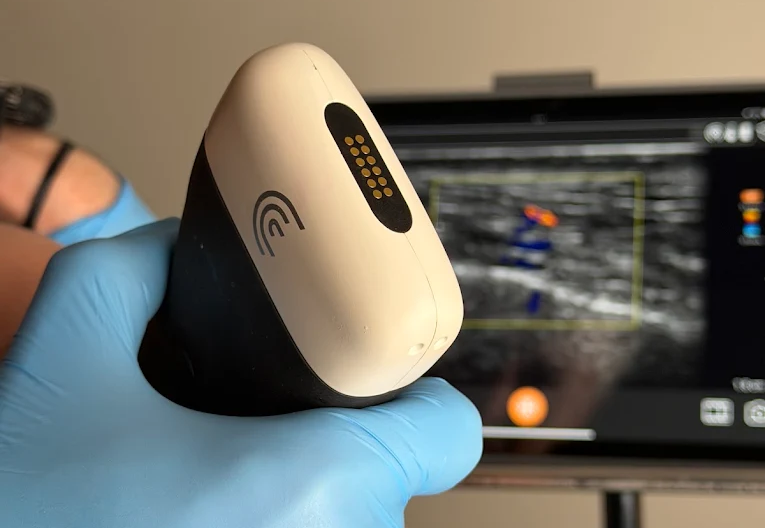
When most people think about ultrasound, they think about pregnancy, abdominal imaging, or examining muscles and joints.
But high-frequency ultrasound is increasingly being used in aesthetic medicine to look beneath the surface of the face.
At Nuvel Skin & Laser, we have ultrasound available as an additional tool when assessing and treating patients with dermal fillers.
It can provide information that simply isn’t available by looking at or feeling the face alone.
But it’s important to understand what ultrasound can-and cannot-do.
Why would we use ultrasound for Facial Injectables?
The face contains a complex network of arteries, veins, muscles, fat compartments, glands, and previously placed injectable products.
We have detailed anatomical knowledge of where these structures are typically located.
The important word, however, is typically.
Anatomy varies from person to person.
Ultrasound allows us, in selected situations, to look beneath the skin and obtain additional information about an individual patient’s anatomy.
Seeing Blood Vessels Beneath the Skin
One of the most useful applications of ultrasound in aesthetic medicine is identifying blood vessels.
Using Doppler ultrasound, blood flow can be visualized in real time.
This can help identify the location and depth of vessels in an area being assessed or treated.
This is particularly valuable because facial vascular anatomy isn’t identical in every person. A vessel may not always be exactly where an anatomical diagram suggests it should be.
Ultrasound therefore adds another layer of information to our understanding of facial anatomy.
Finding Previous Filler
Patients don’t always know exactly what filler they’ve had, where it was placed, or even when it was injected.
Sometimes treatment was performed years earlier.
High-frequency ultrasound can often help identify previously placed filler and provide information about where it sits within the tissues.
This can be useful when assessing:
- Persistent swelling
- Lumps or nodules
- Unexpected facial contours
- Previous filler before additional treatment
- Areas where the patient’s treatment history is uncertain
This is becoming increasingly relevant as we learn that some fillers may remain detectable considerably longer than patients were historically led to expect.
Ultrasound and Filler Complications
Ultrasound can also be useful when investigating certain dermal filler complications.
For example, it may help identify the location of filler, surrounding tissue changes, and relevant vascular structures.
In selected circumstances, ultrasound can also assist with targeted treatment when filler needs to be dissolved.
This can be particularly useful when the exact location of previously injected hyaluronic acid filler isn’t obvious from examining the surface of the face.
Can Ultrasound Prevent Every Vascular Occlusion?
No.
This is an important distinction.
Ultrasound is a valuable tool, but it does not make dermal filler treatment risk-free.
Blood vessels are small and three-dimensional. Anatomy varies. The face moves. Injection itself changes tissue position, and it isn’t practical or necessary to scan every millimetre of every routine filler treatment.
Ultrasound should therefore not be viewed as a replacement for:
- Detailed anatomical knowledge
- Appropriate injection technique
- Careful product selection
- Understanding appropriate tissue planes
- Recognition of complications
- Clinical judgment
It is another layer of information—not a guarantee.
Do We Ultrasound Every Patient Before Filler?
No.
Many routine injectable treatments can be safely planned and performed based on clinical assessment, anatomical knowledge, and appropriate technique.
We use ultrasound when we believe the additional information is useful.
That may include evaluating an anatomical area before treatment, locating a vessel, investigating previous filler, or assessing a potential complication.
Having the technology available means we can use it when the clinical situation warrants it rather than pretending every patient requires an ultrasound examination.
Technology Doesn’t Replace Judgment
It’s easy to assume that more technology automatically means safer or better treatment.
That’s not necessarily true.
An ultrasound image is only useful if the person using it understands what they’re looking at and how that information relates to the treatment being considered.
The same principle applies to injectables themselves.
A syringe of filler is simply a tool.
So is ultrasound.
Neither replaces assessment, anatomical knowledge, experience, or judgment.
Why We Use Ultrasound at Nuvel Skin & Laser
At Nuvel Skin & Laser, our approach to injectable treatment is based on understanding the face before deciding how to treat it.
High-frequency ultrasound gives us another way to gather information when needed.
It can help us:
- Visualize vascular anatomy
- Assess the depth and location of certain structures
- Identify previously placed filler
- Investigate some filler-related concerns
- Assist with management when complications occur
For us, ultrasound isn’t about making dramatic claims that injectable treatment can be made completely risk-free.
It’s about having another useful tool available when more information can help us make a better clinical decision.
A Physician-Led Approach to Dermal Fillers in Vernon, BC
Dermal fillers are medical procedures.
At Nuvel Skin & Laser in Vernon, BC, treatment begins with an individualized facial assessment and a discussion about your goals, anatomy, treatment options, and potential risks.
When ultrasound can provide useful additional information, we have the ability to incorporate it into that assessment or treatment.
The goal remains the same whether ultrasound is needed or not: thoughtful treatment, appropriate planning, and natural-looking results.
If you’re considering dermal fillers in Vernon, BC, book a consultation with Nuvel Skin & Laser to discuss whether treatment is appropriate for you.